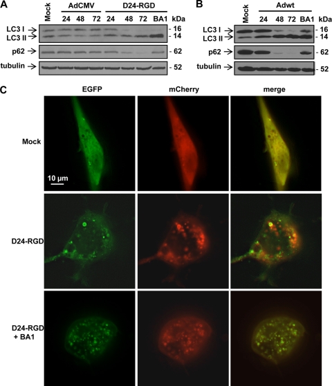
Fig. 2.
Adenovirus induces a complete autophagic flux. (A) U-87 MG cells were infected with replication-deficient adenoviral vector AdCMV (10 PFU/cell) and Delta-24-RGD (D24-RGD; 10 PFU/cell). (B) U-251 MG cells were infected with Adwt (10 PFU/cell). Cells were collected at 24, 48, and 72 h after infection. Bafilomycin A1 (BA1; 10 nM) was added to the cells 24 h after viral infection, and cells were collected 48 h later. Cell lysates were subjected to immunoblot analysis. Tubulin expression was used as a loading control. (C) Representative images of mCherry-EGFP-LC3 fluorescence in the U-87-MG cells. Cells were first transfected with plasmid expressing mCherry-EGFP-LC3. Twenty-four hours later, cells were infected with Delta-24-RGD at 50 PFU/cell in the absence or presence of 10 nM bafilomycin A1. After 48 h, live cells were observed under fluorescence microscopy.

