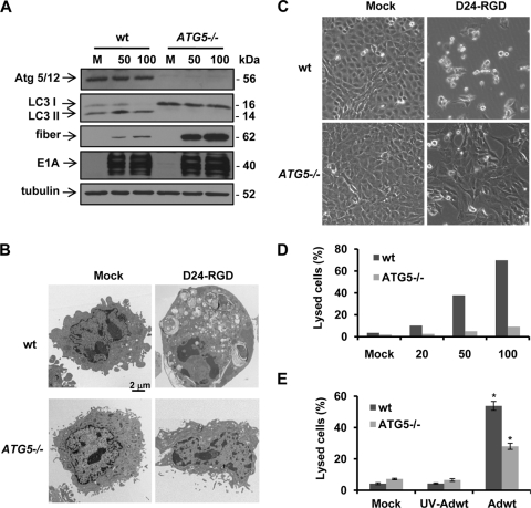
Fig. 5.
ATG5 knockout blocks adenovirus-induced autophagy and cell lysis in wild-type (wt) or ATG5 knockout (ATG5−/−) MEFs. (A) Cells were infected with Delta-24-RGD at the indicated doses (PFU/cell). After 72 h, cell lysates were collected for immunoblot analysis. Tubulin expression was used as a loading control. (B) Representative electron micrographs. Cells were infected with Delta-24-RGD at 50 PFU/cell for 72 h. Note that numerous vacuoles are induced by the virus in wt MEFs but only a few are induced in ATG5−/− MEFs. (C) Phase-contrast images of MEFs infected with Delta-24-RGD at 100 PFU/cell for 72 h. (D) Cell lysis caused by Delta-24-RGD in MEFs. Cells were infected with Delta-24-RGD at the indicated doses (PFU/cell). Seventy-two hours later, the cells were assayed for cell lysis. (E) Cells were infected with Adwt at 100 PFU/cell. Seventy-two hours later, the cells were assayed for cell lysis. Three independent experiments were performed. Data are shown as means ± SD. *, P = 0.004. M; mock; D24-RGD, Delta-24-RGD; UV-Adwt, UV-inactivated Adwt.

