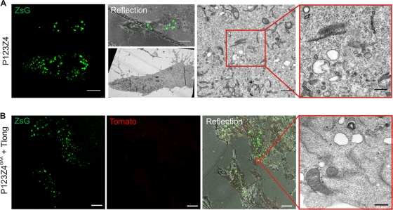
Fig. 8.
Requirements for spherule formation. BSR cells were transfected with the polyprotein P123Z4 alone (A) or with polymerase-defective P123Z4GAA together with the template Tlong (B). The sample was fixed at 24 h posttransfection, imaged with a confocal microscope, and then prepared for CLEM. Representative images are shown. In the EM images of transfection-positive cells, intracellular vacuoles (A) or plasma membrane (B) devoid of spherules can be seen. Bars, rightmost EM images, 200 nm; confocal microscopic images, 10 μm.

