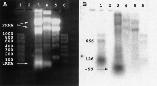
Figure 5.
Northern blot detection of Ciona SL RNA. (A) Fluorescence of ethidium bromide stained gel. (B) Autoradiography following transfer to nylon membrane and hybridization with a 5′-32P-labeled oligonucleotide complementary to the SL sequence. (Lanes 1,6) An RNA marker set (100–1000 nucleotide sizes indicated), to which has been added either 50 ng (lane 1) or 5 ng (lane 6) each of 126-nucleotide, and 666-nucleotide SL-containing in vitro transcripts of a plasmid encoding mRNA 3 (see Fig 2). (Lane 2) Blank; (lane 3) Ciona body-wall muscle RNA (not salt precipitated); (lane 4) quail muscle RNA (not salt precipitated); (lane 5) Ciona body-wall muscle RNA (salt precipitated). Large and small subunit rRNA and tRNA bands are indicated in A. Because the samples had not been salt precipitated, lanes 3 and 4 also contain genomic DNA (near sample wells).

