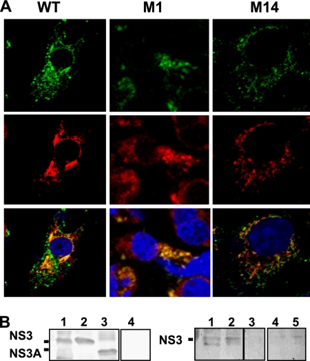
Fig. 3.
Interaction of BTV NS3 with S100A10/p11 in infected mammalian cells. (A) Colocalization of NS3 (green) with S100A10/p11 (red). BSR cells were infected with WT or mutant viruses as indicated and at 24 h postinfection were fixed, permeabilized, and processed for confocal microscopy. (B) Coimmunoprecipitation of NS3 and S100A10/p11. BSR cells were infected with WT virus (lane 1) or mutant viruses BTVM1 (lane 2) and BTV M14 (lane 3) and at 36 h postinfection were harvested and processed as described in Materials and Methods. Expression of NS3 in cell lysates was assayed by Western blotting using an antibody against NS3 (left panel). As control, a lysate from noninfected cells (lane 4) was included. The coimmunoprecipitation of NS3 WT virus (right panel, lane 1) or mutants M1 (lane 2) and M14 (lane 3) with S100A10/p11 was performed using a monoclonal antibody that recognized S100A10/p11 and analyzed by Western blotting with an antibody against NS3. As controls, a lysate from mock-infected cells (lane 4) and a lysate from cells infected with WT virus but without antibody against S100A10/p11 (lane 5) were included.

