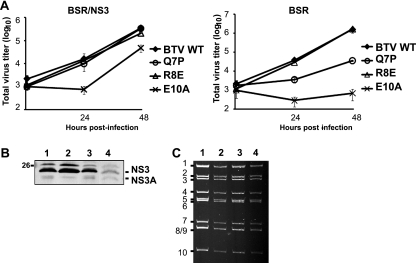
Fig. 5.
Characterization of mutant BTVQ7P, BTVR8E, and BTVE10A viruses. (A) Complementary BSR/NS3 cells (left panel) or normal BSR cells (right panel) were infected with mutant virus BTV Q7P, R8E, or E10A or with the WT and harvested at different time points postinfection as indicated. Total titer was determined by plaque assay, expressed as PFU/ml, and plotted on a logarithmic scale. (B) The expression of NS3 was assessed by Western blotting in BSR cell lysates processed at 48 h postinfection with mutant virus Q7P (lane 1), R8E (lane 2), or E10A (lane 3). Lysate from cells infected with WT BTV (lane 4) was included as control. The number on the left indicate the molecular mass standard in kDa. (C) Genomic dsRNA purified from BSR cells infected with BTVQ7P (lane 2), BTVR8E (lane 3), or BTVE10A (lane 4) was purified and analyzed in a nondenaturing polyacrylamide gel. As a control, dsRNA from WT virus was included (lane 1).

