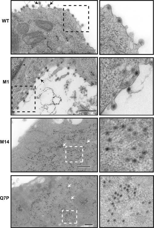
Fig. 7.
Ultrastructural analysis of mammalian cells infected with mutant or WT viruses. BSR cells were infected with WT, BTVM1, BTVM14, or BTVQ7P virus, and at 24 h postinfection cells were processed for sectioning. Arrows in the left panels indicate particles. An area of each section is amplified in the right panels to show the detail. Bars, 500 nm.

