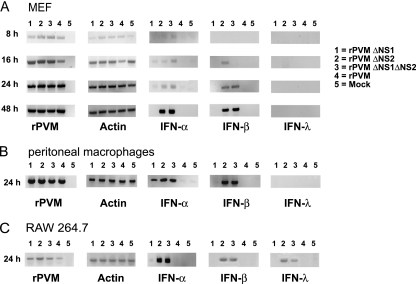
Fig. 2.
Induction of IFN-α, IFN-β, and IFN-λ in infected cell cultures, as detected by RT-PCR. MEF (A), peritoneal macrophages of C57BL/6 mice (B), or the RAW 264.7 macrophage cell line (C) were either mock infected or infected with rPVM or the ΔNS1, ΔNS2, or ΔNS1ΔNS2 mutant at an input MOI of 1 PFU per cell. At the indicated time points, total cellular RNA was extracted, and 1 μg of total RNA was subjected to RT-PCR to amplify mRNA specific for rPVM N, total actin, IFN-α5, IFN-β, or IFN-λ2/3. The RT-PCR products were subjected to electrophoresis on agarose gels and visualized with ethidium bromide.

