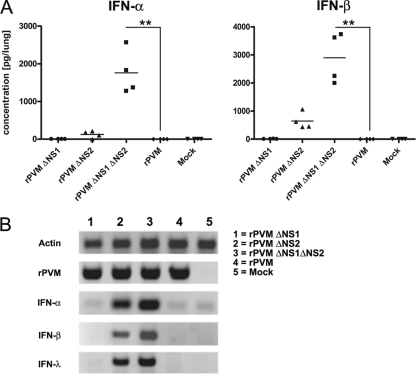
Fig. 4.
Induction of IFN-α, IFN-β, and IFN-λ in the lungs of infected mice. C57BL/6 mice in groups of four were mock infected or infected intranasally with 50,000 PFU of rPVM or the ΔNS1, ΔNS2, or ΔNS1ΔNS2 mutant. The animals were sacrificed after 24 h, and the lungs were removed, homogenized, and aliquoted. (A) One aliquot was used to determine IFN-α and -β protein levels by ELISA. Statistical significance was calculated by analysis of variance, followed by Tukey-Kramer posthoc test analysis. Columns marked with asterisks differ significantly (*, P ≤ 0.05; **, P ≤ 0.01; ***, P ≤ 0.001) from values for rPVM. Data from one of two independent experiments providing comparable results are shown. (B) Total RNA was extracted from a second aliquot of lung homogenate and IFN-α5-, -β-, and -λ2/3-specific mRNA, as well as PVM- and actin-specific mRNA, was amplified by RT-PCR and visualized by gel electrophoresis and ethidium bromide staining. Shown are exemplary samples of one mouse per infection group.

