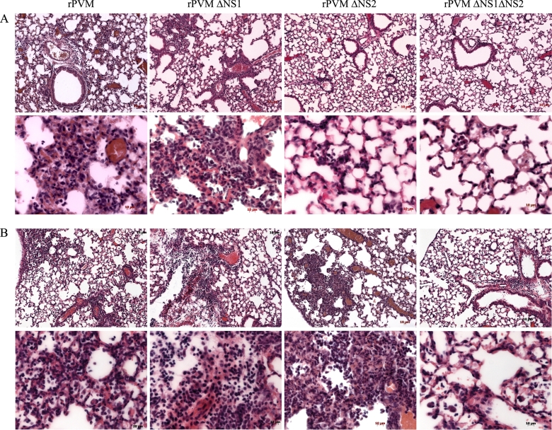
Fig. 7.
Histopathology of C57BL/6 and B6.IFNAR0/0.IL28Rα0/0 mice after infection with rPVM or the NS deletion mutants. Histopathological analysis of lung sections (H&E staining) from C57BL/6 (A) and B6.IFNAR0/0.IL28Rα0/0 (B) mice that had been infected with 250 PFU of rPVM, 5,000 PFU of rPVM ΔNS1, or 50,000 PFU of the ΔNS2 and ΔNS1ΔNS2 mutants 8 days previously. The panels show representative sections from histological analysis of two to four mice per group. Scale bars are indicated in the panels.

