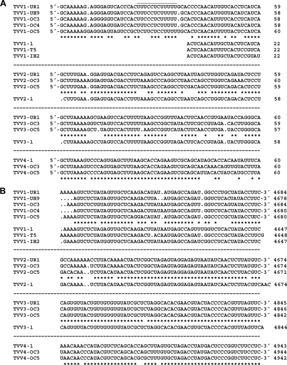
Fig. 4.
Terminal sequences of TVV plus strands. Sequences at the 5′ (A) and 3′ (B) termini are shown. Multiple sequence alignments for the viruses within each species (TVV1, -2, -3, and -4) were performed by the program Clustal 2.0.12, with default settings, at http://www.ebi.ac.uk/Tools/clustalw2. Gaps within each sequence are indicated by periods. Conserved positions in the consensus are indicated by asterisks below each set of newly determined sequences. Previously reported TVV sequences are shown below the consensus in each set. The positions of the rightmost nucleotide in each line of sequence are indicated at right.

