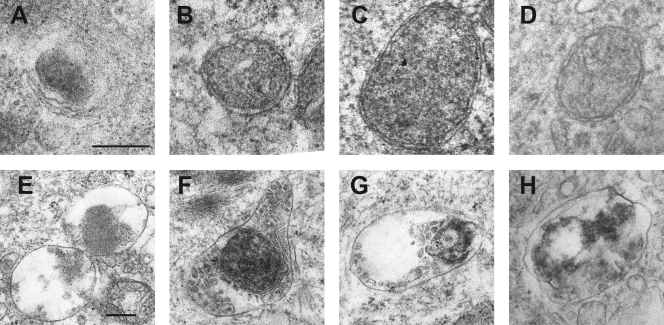
Fig. 5.
Autophagy-related structures in HCMV-infected HFFF2 cells. Images from the electron microscopic examination of HFFF2 cells infected with gradient-purified HCMV or UV-HCMV for 6 h. (A) Possible engulfment by a phagophore structure; (B to D) double-membraned autophagosomes; (E to H) autolysosomes with cargo in different stages of digestion. The bar in panel A represents 200 nM for panels A to D, and the bar in E represents 200 nM for panels E to H.

