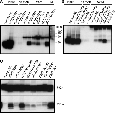
Fig. 2.
Immunoprecipitation of PrPSc from CJD samples with MAb W261. (A) Detection of PrPSc in various sporadic (s) cases and one genetic (g) case of CJD. (B) Detection of PrPSc in various CJD cases, including vCJD. (C) Demonstration of various amounts of PrPSc in spontaneous, genetic, and variant CJD cases tested in panels A and B by Western blotting. Immunoprecipitations were performed using 1 ml of 1% brain homogenate, and complete supernatants of boiled precipitates were analyzed by Western blotting. For input material, 30 μl of 1% brain homogenate was loaded per lane. The signal at 50 kDa corresponds to the heavy chain of mouse immunoglobulin.

