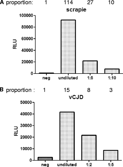
Fig. 3.
ELISA with MAb W261 as the capture antibody for the detection of PrPSc. (A) Detection of sheep PrPSc; (B) detection of vCJD PrPSc. The specificity of this capture ELISA is demonstrated for sheep scrapie (A) and for vCJD (B). For each species, a control homogenate from normal brains was only weakly recognized, whereas the homogenate from scrapie- or vCJD-affected brains was clearly detected in a dose-dependent manner. In conclusion, this sandwich ELISA using MAb W261 is able to distinguish between TSE-infected and uninfected tissue homogenates of sheep or humans without the need for proteinase K digestion, demonstrating its usefulness for diagnostic assays. Negative controls (neg) are uninfected sheep or human brain homogenates. One microliter of 10% or further diluted brain homogenate was used per sample in a volume of 200 μl. The results are given as relative light units (RLU). The proportions above the graphs represent the values obtained divided by the value for the negative control.

