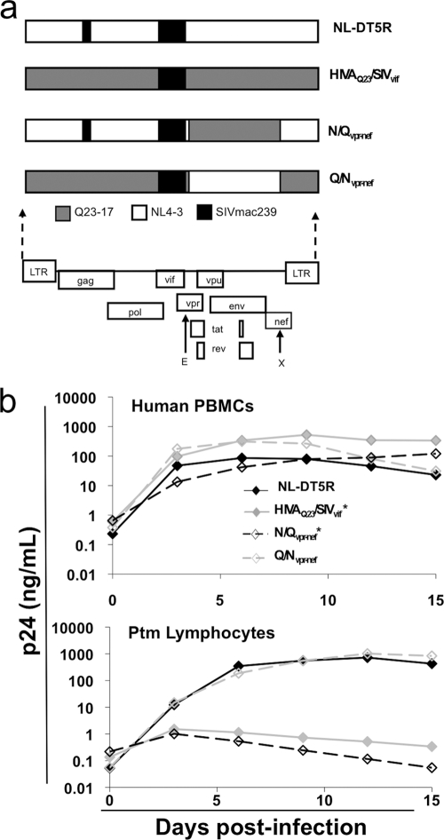
Fig. 1.
Infection of human PBMCs and Ptm lymphocytes with minimal SHIVs. (a) Schematic representation of the contribution of Q23-17, NL4-3, and SIVmac239 sequences to the minimal SHIVs used for replication experiments. E and X represent the EcoRI and XhoI sites used to make N/Qvpr-nef and Q/Nvpr-nef. (b) p24gag levels shown as a function of time postinfection in human PBMCs and Ptm lymphocytes. The data points represent the average measurements from duplicate infected cultures. The figure key is shown in the top plot, and viruses with an asterisk were used at a 10-fold-higher MOI for infection of Ptm cells. The results are representative of at least three independent experiments.

