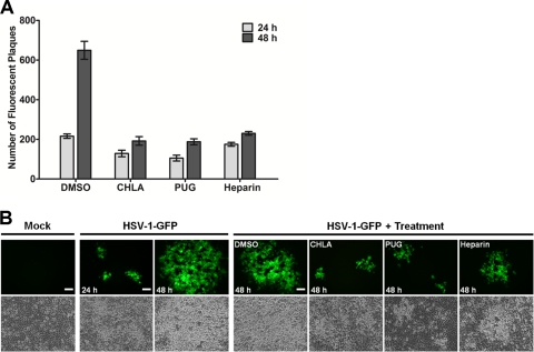
Fig. 7.
CHLA and PUG can limit HSV-1 secondary infection and cell-to-cell spread of the virus. A549 cells were infected with HSV-1-GFP (200 PFU/well) for 1 h and then treated with citrate buffer (pH 3.0) to inactivate noninternalized extracellular viral particles. Cells were overlaid with medium alone (A) or medium containing 0.1% neutralizing antibody (B). After a p.i. incubation period of 12 h (A) or 24 h (B), the infected cells were treated with CHLA (60 μM), PUG (40 μM), heparin (100 μg/ml), or DMSO (0.1%), before further incubation for a total of 48 h after the initial infection. Over the course of infection subsequent to the drug addition, the plates were scanned and quantified for fluorescent viral plaques (A) or photographed using an inverted fluorescence microscope at ×100 magnification (B). (A) Number of fluorescent plaques counted between 24 and 48 h p.i. with drug treatment initiated at 12 h after viral challenge. The data shown are means ± the SEM of three independent experiments with each treatment performed in duplicate. (B) Comparison of viral plaque size between 24 and 48 h p.i. with drug treatment initiated at 24 h after viral challenge. Scale bars, 100 μm. Representative images are from one of two independent experiments.

