Fig. 8.
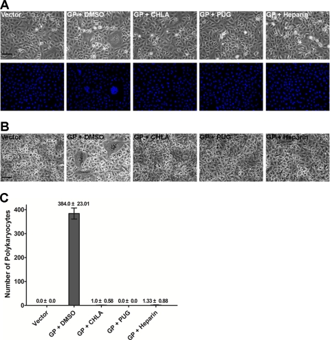
CHLA and PUG can prevent HSV-1 glycoprotein-mediated cell fusion events. A549 cells were transfected with plasmids expressing the individual HSV-1 glycoproteins (gB, gD, gH, and gL). After 6 h of transfection, cells were washed with PBS and treated with CHLA (60 μM), PUG (40 μM), heparin (100 μg/ml), or DMSO (0.1%). After further incubation for 24 h, cells were fixed with methanol and stained with Hoechst dye (A) or Giemsa (B). Photomicrographs were then taken at ×200 magnification. (A) Phases (upper panels) and respective fluorescence pictures displaying the Hoechst dye-stained nuclei (bottom panels). (B) Giemsa-stained cells in a similar experiment. Representative pictures shown are from one of three independent experiments. Vector, empty vector; GP, HSV-1 glycoproteins; scale bars, 100 μm. (C) The total number of polykaryocytes (>10 nuclei) from each treatment was quantified. The data shown are means ± the SEM of three independent experiments.

