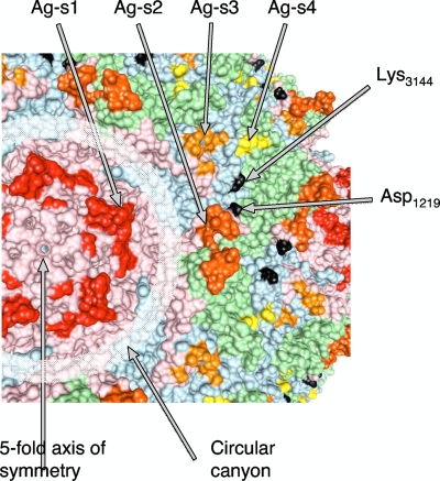
Fig. 3.
Location of the epitope to antibody H2 on the surface of type 1 poliovirus. Capsid proteins VP1, VP2, and VP3 are shown in pink, green, and blue, respectively. Antigenic sites 1, 2, 3, and 4 are shown in red, orange, gold, and yellow, respectively. Amino acids that are replaced in escape mutants are shown in black.

