Fig. 2.
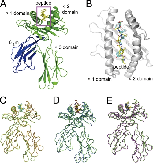
Overview of the structure of bovine MHC I N*01801. (A) Overview of the structure of the bovine MHC I N*01801 represented by molecule 1 in the asymmetric unit. (B) The peptides (yellow for the peptide of molecule 1 and cyan for the peptide of molecule 2) are presented in the peptide-binding clefts. (C) Superposition of the two molecules with one asymmetric unit of N*01801 (green for molecule 1 and orange for molecule 2). (D) Structural superposition of the bovine MHC I N*01801 molecule 1 (green) with N*01301 (blue) (PDB code 2XFX). (E) Structural alignments of the MHC heavy chain and β2m show the similar overview conformations of N*01801 molecule 1 (green) and HLA-B*5101 (purple) (PDB code 1E27). This figure and subsequent figures showing molecule models were generated using PyMol (www.pymol.org).

