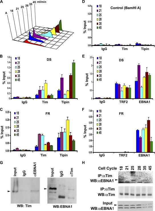
Fig. 2.
Cell-cycle-dependent association of Tim and Tipin at OriP. (A) Mutu I cells were fractionated by centrifugal elutriation and then assayed by FACS. Elutriation fractions are indicated in ml/min. (B) ChIP assay with anti-Tim, anti-Tipin, or control IgG at the DS region for each stage of the cell cycle as indicated. (C) Same as in panel B, except for analysis at the FR region. (D) Same as in panel B, except for analysis at the control BamHI A region. (E) ChIP assay with anti-EBNA1, anti-TRF2, or control IgG at the OriP DS region for each stage of the cell cycle, as indicated. (F) Same as in panel E, except for analysis at the FR region. (G) Co-IP analysis of EBNA1 and Tim in asynchronous Mutu I cells. EBNA1 or control IgG IPs were assayed by Western blotting (WB) for Tim protein (left panel). Tim or control IgG IPs were assayed by Western blotting for EBNA1 protein (right panel). Arrowheads indicate the expected sizes of the indicated proteins. αEBNA1, anti-EBNA1; αTim, anti-Tim. (H) Cell cycle fractions from Mutu I cells were subject to IP with antibody to Tim and then assayed by Western blotting with anti-EBNA1 (top panel) or anti-Tim (middle panel). The EBNA1 input for each cell cycle fraction is shown in the lower panel.

