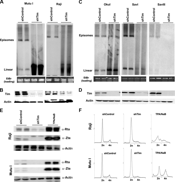
Fig. 4.
Loss of EBV episomal forms in Burkitt's lymphoma cell lines after Tim depletion. (A) Pulsed-field electrophoresis gel analysis of Mutu I (left) or Raji (right) cells after infection with shControl or shTim lentivirus. EBV DNA was detected by Southern blotting. Episomes and linear forms of EBV genomes are indicated. EtBr, ethidium bromide. (B) Western blot of cells used for pulsed-field analysis shown in panel A. Antibodies for Tim (top panel) or actin (lower panel) are indicated. (C) Same analysis as described in panel A for Oku I, Sav I, and Sav III cells. (D) Western blots showing Tim (top) and actin (lower) levels for cells analyzed in panel C. (E) Western blot analysis of EBV lytic proteins Rta, Zta, and control actin for Raji and Mutu I cells after infection with shControl or shTim, or after treatment with tetradecanoyl phorbol acetate (TPA) and NaB for 48 h, as indicated. α-, anti-. (F) FACS analysis of cell cycle profiles for Raji and Mutu I cells after infection with shControl or shTim or after treatment with TPA and NaB for 48 h.

