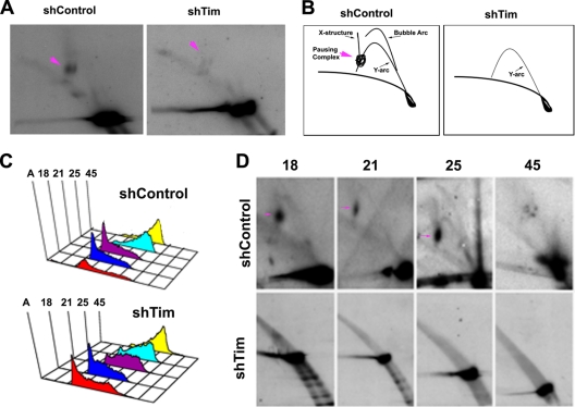
Fig. 5.
Tim depletion causes a loss of replication fork structures at OriP. (A) 2D neutral agarose gel electrophoresis and Southern blot analysis of OriP DNA in Mutu I cells after infection with shControl (left panel) or shTim (right panel) lentivirus. Pink arrows indicate replication fork pausing structures. (B) Schematic interpretation of 2D neutral agarose gels shown in panel A. (C) FACS analysis of cell cycle fractions from centrifugal elutriation after shControl lentivirus infection of Mutu I cells. (B) Same as in panel A, except for shTim lentivirus infection. (D) 2D neutral agarose gel analysis of OriP DNA from shControl (top row)- or shTim (bottom row)-infected Mutu I cells. Cell cycle fractions 18, 21, 25, and 45, are indicated. Replication fork pausing structures are indicated by pink arrows.

