Fig. 6.
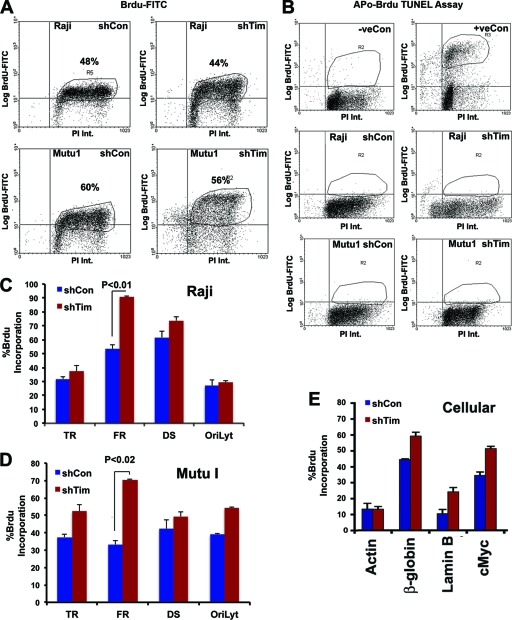
Accelerated DNA replication through FR in Tim-depleted cells. (A) FACS analysis of Raji (top panels) or Mutu I (lower panels) cells infected with shControl (left panels) or shTim (right panels) after pulse-labeling with BrdU for 30 min, followed by staining with propidium iodide (PI). BrdU intensity (Int.) was monitored by anti-BrdU-conjugated fluorescein isothiocyanate (FITC) (y axis), and PI was monitored on the x axis. (B) Apo-BrdU TUNEL assay for Raji (middle) or Mutu I (lower) after shControl (left) or shTim (right) infection. Control samples were camptothecin-treated (+) or untreated (−) HL60 cells provided by the manufacturer (top panels). (C) BrdU-ChIP assay for Raji cells after infection with shControl (blue) or shTim (red). BrdU incorporation was assayed at EBV locations for terminal repeats (TR), FR, DS, and OriLyt, as indicated. (D) Same as in panel C, except Mutu I cells were analyzed. (E) Same as in panel D, except BrdU was assayed at the cellular loci for actin, β-globin, lamin B, and c-Myc in Mutu I cells. Error bars indicate standard deviations from the mean, and P values were determined by Chi-square test.

