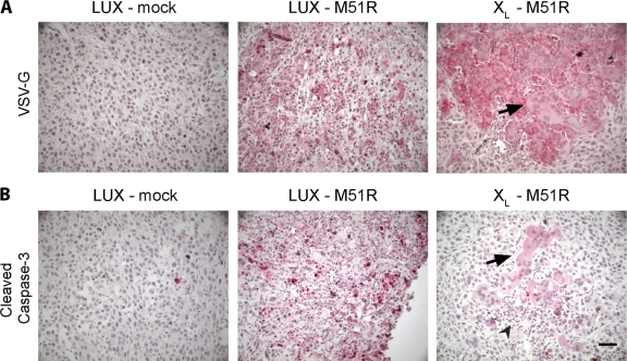
Fig. 7.
Expression of viral antigen and cleaved caspase-3 in control and Bcl-XL-overexpressing tumors infected with M51R VSV in vivo. (A) Representative micrographs of tumor sections immunostained for VSV glycoprotein (VSV-G). Red indicates positive immunoreactivity. Large syncytia were seen in infected Bcl-XL tumor cells (black arrow). (B) Tumor sections were stained for cleaved caspase-3. Low to moderate staining in Bcl-XL tumor cells was only seen where syncytia had formed (black arrow). Immune infiltrating cells could be clearly identified at the site of syncytium formation (arrowhead). Bar, 30 μm.

