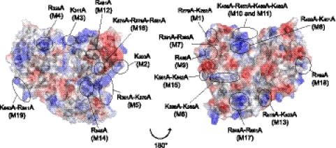
Fig. 3.
Amino acid positions of mutations introduced in the RdRp domain of NS5. A “back” view of the structure of the DENV3 RdRp domain (Protein Data Bank [PDB] accession code 2J7U) in surface representation (left) and a “front” view of the structure of the DENV3 RdRp domain rotated 180° (right) are shown. Surfaces are represented with the electrostatic potential in blue for positive charges and in red for negative charges. The figure was drawn using the PyMOL program.

