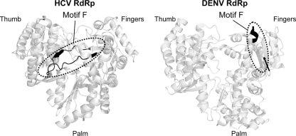
Fig. 8.
Position of the motif F on HCV and DENV RdRps. Structure of HCV (PDB 1NB6) and DENV (PDB 2J7U) RdRps (back view) in ribbon representation. The position of the F motif is indicated in black; for DENV RdRp the region of motifs F1 and F2 is missing in the structural model (from amino acids 454 to 466). The position of amino acid 453 is indicated by an arrow. The figure was drawn using the PyMOL program.

