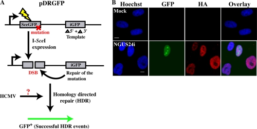
Fig. 1.
Efficient expression of I-SceI from an Ad-based system in HFFs with an integrated pDRGFP substrate. (A) Schematic diagram of reporter plasmid pDRGFP, depicting I-SceI-induced DSB-initiated HDR to produce functional GFP. (B) Representative IF staining of mock- or NGUS24i-infected clone 4. Cells were seeded onto glass coverslips and mock or Ad infected. Coverslips were harvested and processed at 72 hpi as described previously (27). GFP+ cells were scored as HDR competent. Cells were also stained with HA-specific Ab (mouse monoclonal antibody [MAb; IgG2b] 12CA5; Abcam) to detect HA-I-SceI. In all IF experiments, isotype-specific secondary Abs conjugated with either tetramethyl rhodamine isothiocyanate (TRITC; Jackson ImmunoResearch Laboratories) or Alexa Fluor 488 (Molecular Probes) were used to detect mouse primary Abs. Nuclei were counterstained in all experiments with Hoechst dye. Scale bar, 5 μm for all panels in all figures.

