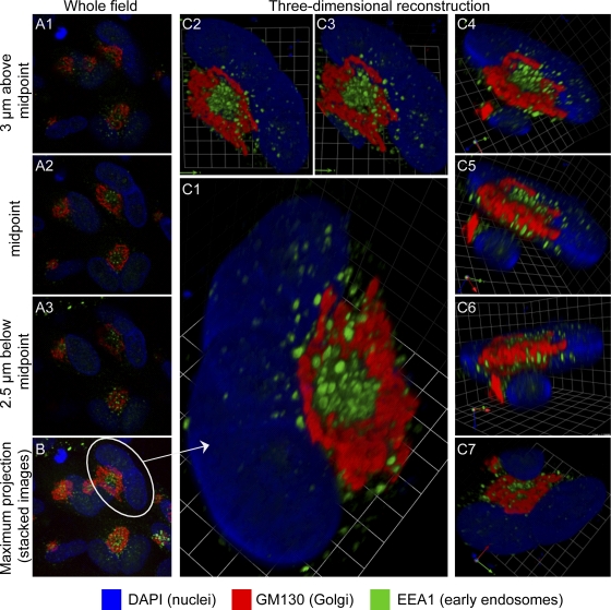
Fig. 1.
Relationships between images from single confocal planes and reconstructed 3-D images. HCMV(AD169)-infected lung fibroblasts (HLF cells) were stained for the indicated markers at 120 hpi. Serial 0.5-μm confocal sections were obtained with a Leica TCS SP5 laser-scanning confocal microscope. (A1 to A3) Three of the 23 confocal sections that were used to generate the 3-D reconstruction shown in panel C. The location of each section in the Z-series is indicated. (B) Maximum projection image of the entire field containing the cell from which the 3-D reconstruction was made. This shows that many infected cells have related structures. (C to C7) A collection of views of a 3-D reconstruction of a single binucleated infected cell (circled in panel B). A portion of the nucleus of an overlapping adjacent cell is visible in panels C4 through C7. Panel C7 is a view from the bottom. Grid spacing is 3.31 μm. A video showing the reconstruction rotating in space is available as Video S1 in the supplemental material.

