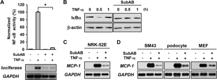
Fig. 1.
Suppression of TNF-α-induced activation of NF-κB by SubAB. (A) NRK/NFκB-Luc cells were pretreated with or without 10 ng/ml SubAB for 24 h, exposed to 10 ng/ml TNF-α for 6 h, and subjected to a luciferase assay (top) and Northern blot analysis of luciferase mRNA (bottom). Activity of luciferase was normalized by the number of viable cells estimated by formazan assay, and relative values (%) are shown. Reporter assay and formazan assay were performed in quadruplicate. Data are presented as means ± SE. Statistical analysis was performed using the nonparametric Mann-Whitney U test to compare data in different groups. An asterisk indicates a statistically significant difference (P < 0.05). (B) NRK-52E cells were pretreated with or without SubAB, exposed to TNF-α for up to 1 h, and subjected to Western blot analysis of IκBα. The level of β-actin is shown at the bottom as a loading control. (C and D) NRK-52E cells, SM43 cells, podocytes, and MEF were pretreated with or without SubAB, exposed to TNF-α for 6 h, and subjected to Northern blot analysis of MCP-1. Expression of GAPDH is shown at the bottom as a loading control.

