Fig. 3.
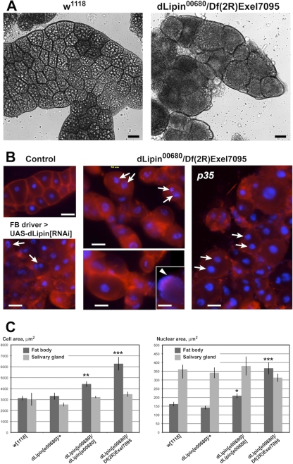
Loss of dLipin causes defects in fat body development. (A and B) The size and shape of fat body cells is dramatically altered in dLipin-deficient larvae. (A) Bright-field microscopy indicates that, compared to wild-type cells (w1118), fat body cells of third-instar larvae lacking dLipin are more variable in size, more rounded, and, in some regions, detached from one another. Large fat droplets that appear as bright inclusions, and are plentiful in the control fat body, are scarce in the fat body lacking dLipin. (B) Staining with the plasma membrane stain CellMask (red) highlights cell boundaries, confirming the cell shape changes, while staining with the DNA stain DAPI (blue) reveals that cell nuclei in some of the dLipin-deficient cells are fragmented (arrows). The arrowhead marks a cell in which the DNA is dispersed throughout the cell. Cells show a similar mutant phenotype in the presence of the apoptosis inhibitor p35 or after dLipin knockdown by RNAi. Bars, 50 μm. (C) The sizes of cells and nuclei are increased in the fat bodies, but not in the salivary glands, of dLipin mutants, indicating that the observed effects are specific for fat tissue. Asterisks indicate a significant difference from the mean for w1118 flies (*, P < 0.05; **, P < 0.01; ***, P < 0.0001). Error bars indicate standard errors of the means.

