Fig. 4.
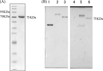
The S. ruminantium flagellin is glycosylated. (A) SDS gel pattern of an S. ruminantium flagellin. First lane, molecular size marker; second lane, single band of flagellin. (B) Detection of glycosylation of an S. ruminantium flagellin (lanes 3 and 6). Salmonella SJW1103 flagellin was used as a negative control (lanes 1 and 4), and Azospirillum flagellin was used as a positive control (lanes 2 and 5). Coomassie brilliant blue (CBB) staining (left) and PAS staining (right) of the purified flagellins are shown.

