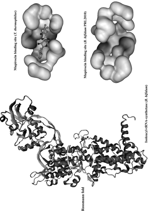
Fig. 3.
Predicted 3D structure of the isoleucyl-tRNA synthetase from B. bifidum PRL2010 obtained by homology modeling using T. thermophilus as the template. This image is based on a docking model. The magnified sections represent the active site characterized by a Rossmann-type folding of the isoleucyl-tRNA synthetase from T. thermophilus as retrieved from the PDB and interacting with the mupirocin molecule, as well as the homologous region of the isoleucyl-tRNA synthetase from B. bifidum PRL2010. These structures are depicted as ball-and-stick structures shaded according to atom type. The different levels of interaction with the IleS binding cavity are indicated by various levels of gray shading.

