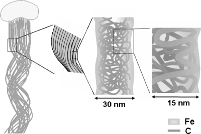
Fig. 4.
Schematic of the putative carbon fibril network comprising a single fiber (approximately 30-nm width) of the stalk. Fibrils of a few nanometers were intermingled and folded at the fiber core region but not at the extreme margin. These C fibrils were excreted from the export pores located in the wall region of the bacterial cell. Oxidized iron could interact with C in the entire fiber.

