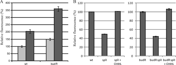
Fig. 5.
(A) Relative expression of plasmid-borne PbudR-gfp in wild-type RVH1 and RVH1 budR in LB medium at pH 7.0 (light gray) or pH 5.5 in the presence of 15 mM acetate (dark gray). Stationary-phase cultures were diluted 1/100 and exposed for 4 h to the indicated conditions before GFP expression was measured. (B) Relative expression of plasmid-borne PbudR-gfp in wild-type RVH1, RVH1 splI, and RVH1 splI complemented with OHHL (left) and in RVH1 budR, RVH1 splI budR, and RVH1 splI budR complemented with OHHL (right). Stationary-phase cultures were grown under the indicated conditions before GFP expression was measured. In each graph, GFP expression of the control strain (i.e., first bar) was arbitrarily set at 100%. Average values and standard deviations for three independent experiments are shown.

