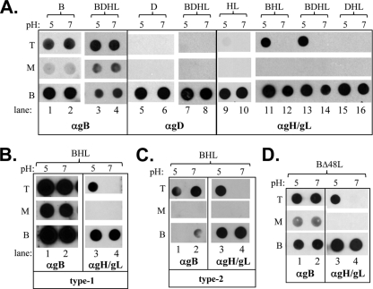
Fig. 1.
(A) Soluble gH/gL cofloats with soluble gB and liposomes upon incubation at low pH. Purified soluble glycoproteins (abbreviated B, D, H, and L) were incubated with liposomes for 1 h at 37°C. Samples were then adjusted to 1 M KCl, incubated for an additional 15 min, and then layered beneath a discontinuous 5 to 40% sucrose gradient. Gradients were centrifuged for 3 h, fractionated, and analyzed by dot blot assay. The top (T), middle (M), and bottom (B) fractions of the gradients are shown. Blots were probed with either the anti-gB PAb R68, anti-gD PAb R8, anti-gH2/gL2 PAb R176, or anti-gD PAb R8. Soluble proteins are all from HSV type 1 (gB1 probed with the type-common PAb R68 and gH1/gL1 probed with PAb R137) (B) or from HSV type-2 (gB2 probed with PAb R68 and gH2/gL2 probed with PAb R176) (C). (D) Flotation was performed with soluble gB and gH2Δ48/gL2 and probed with either R68 or R176. α, anti.

