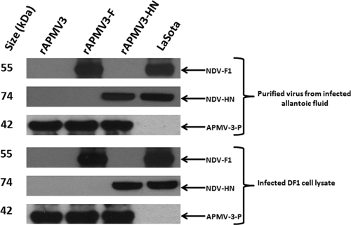
Fig. 2.
Expression of NDV surface proteins F and HN in DF1 cells and their incorporation into rAPMV3 virions. DF1 cells were infected with the individual rAPMV3 constructs, and 48 h later, the cells were collected and processed to prepare cell lysates. In addition, allantoic fluid from embryonated eggs infected with the individual constructs was clarified and subjected to centrifugation on sucrose gradients to make partially purified preparations of virus particles. These samples were analyzed by Western blot analysis using a monoclonal antibody against the HN protein or a rabbit antiserum specific to a C-terminal peptide of the F protein (bottom). In addition, rabbit antisera specific to an N-terminal peptide of the P protein of APMV-3 was also used. Partially purified virus particles of rAPMV3 empty vector (lane 1), rAPMV3-F (lane 2), rAPMV3-HN (lane 3), or rNDV (lane 4) were used in the top three panels. Lysates of DF1 cells that had been infected with rAPMV3 empty vector (lane 1), rAPMV3-F (lane 2), rAPMV3-HN (lane 3), or rNDV (lane 4) were used in the bottom three panels.

