Fig. 3.
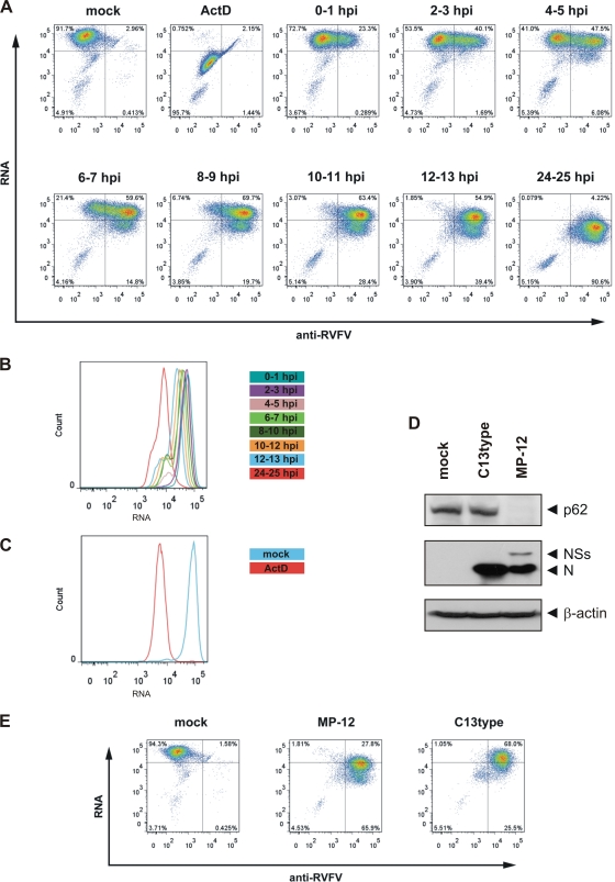
NSs induces host transcriptional suppression. (A) 293 cells were mock infected (mock) or infected with MP-12 at an MOI of 3 and treated with 0.5 mM EU for 1 h at the indicated time points of infection. As a control for transcriptional suppression, cells were treated with 5 μg/ml ActD concurrently with the EU labeling (ActD). Cells were harvested, fixed, and permeabilized immediately after the labeling step. EU-labeled RNA was then detected by click chemistry with an Alexa Fluor 647-coupled azide. The expression of viral proteins was visualized by immunostaining with a polyclonal anti-RVFV antiserum followed by an Alexa Fluor 488-labeled secondary antibody. Cells were then analyzed by flow cytometry. (B) To better demonstrate the progressive loss of transcriptional activity in MP-12-infected cells, data from the experiment whose results are shown in panel A were gated on anti-RVFV-positive cells, and the fluorescence of labeled RNA is displayed as a histogram. (C) Data from the experiment whose results are shown in panel A were gated on anti-RVFV-negative cells, and the fluorescence of labeled RNA in mock-infected and ActD-treated cells is displayed as a histogram. (D) 293 cells were mock infected or infected with either MP-12 or an MP-12 mutant carrying a large deletion in its NSs gene that renders the NSs protein nonfunctional (C13type). Cells were harvested at 12 h.p.i., and whole-cell lysates were analyzed by Western blotting with anti-p62, anti-RVFV, and anti-β-actin antibodies (from top to bottom). (E) 293 cells were mock infected (mock) or infected with either MP-12 or C13type at an MOI of 3 and were treated with 0.5 mM EU from 12 to 13 h.p.i. Cells were harvested, fixed, and permeabilized, and EU-labeled RNA was detected by click chemistry with an Alexa Fluor 647-coupled azide. The expression of viral proteins was visualized by immunostaining with a polyclonal anti-RVFV antiserum followed by an Alexa Fluor 488-labeled secondary antibody. Cells were then analyzed by flow cytometry.

