Fig. 7.
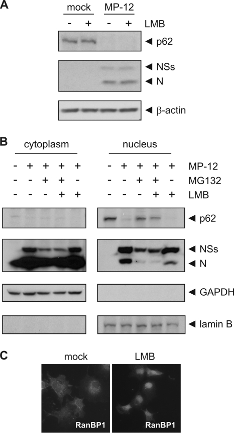
p62 is degraded in the nucleus of infected cells. (A) 293 cells were mock infected or infected with MP-12 at an MOI of 3. Immediately after infection, cells were treated with 20 nM leptomycin B (LMB) where indicated. Cells were harvested at 8 h.p.i., and whole-cell lysates were analyzed by Western blotting with anti-p62, anti-RVFV, and anti-β-actin antibodies (from top to bottom). (B) 293 cells were mock infected or infected with MP-12 at an MOI of 3 and treated with 5 μM MG132 at 3 h.p.i. or 20 nM LMB at 0 h.p.i. where indicated. Cells were harvested at 8 h.p.i. and fractioned into cytoplasmic and nuclear compartments, followed by analysis by Western blotting with anti-p62, anti-RVFV, anti-GAPDH, and anti-lamin B antibodies (from top to bottom). (C) 293 cells were mock treated or treated with 20 nM LMB for 7 h before fixation and permeabilization. RanBP1 localization was visualized by immunostaining. Representative examples of at least 3 independent experiments are shown.

