Fig. 1.
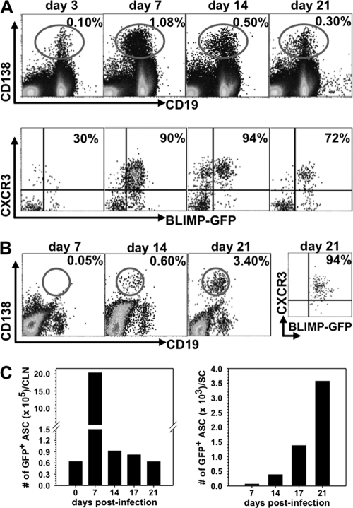
CXCR3+ ASC expand in CLN prior to CNS infiltration. Cells isolated from CLN and spinal cord of naïve and infected BLIMP-1-GFP+/− mice were analyzed for CD19, CD138, GFP, and CXCR3 expression by flow cytometry. (A) Representative plots of CD138 and CD19 expression on CLN cells (top) and of CXCR3 and GFP expression on CD138+ CLN cells (bottom). Numbers depict the percentage of GFP+ CD138+ cells expressing CXCR3. (B) Spinal cord-infiltrating cells were identified by high levels of CD45 (CD45high) surface expression. Dot plots depict CD138+ CD19+ cells within CD45high infiltrates; the far right plot represents the percentage of CD138+ cells expressing both CXCR3 and GFP at day 21 p.i. (C) Changes of total GFP+ CD138+ ASC in the CLN and spinal cord throughout infection. Data are representative of three experiments with pooled organs from 2 to 4 mice/time point/experiment.

