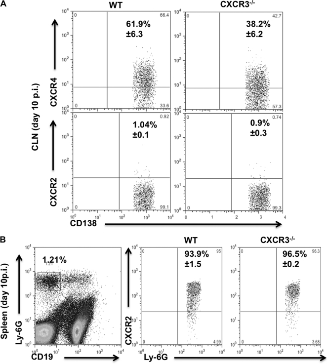
Fig. 6.
ASC do not express CXCR2 but maintain CXCR4 expression even in the absence of CXCR3. Cells isolated from CLN and spleen of JHMV-infected WT and CXCR3−/− mice were analyzed for CD19, CD138, Ly-6G, CXCR4, and CXCR2 expression by flow cytometry at day 10 p.i. (A) Representative plots demonstrating CXCR4, but not CXCR2, expression on ASC. Gates were set on CD138+ CD19lo ASC as depicted by the R1 region in Fig. 2. Numbers depict mean percentages ± standard deviations of ASC expressing CXCR4 or CXCR2, as indicated. (B) CXCR2 staining was validated on splenic neutrophils identified by reactivity with anti Ly-6G MAb. The left plot depicts the percentage of Ly-6G+ CD19− neutrophils in total WT splenocytes at day 10 p.i. The middle and right plots demonstrate CXCR2 expression on cells in the neutrophil gate. Numbers depict mean percentages ± standard deviations of neutrophils expressing CXCR2. In both panels, quadrants were set based on staining with an isotype control MAb. Data are derived from three individual mice per time point.

