Fig. 2.
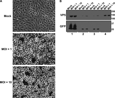
Confirmation of the virion enrichment strategy. (A) Phase-contrast microscopy images of PK15 cells that were either mock infected or infected at an MOI of 1 or 10 at 24 hpi. Scale bar, 20 μm. (B) PK15 cells were either mock infected or infected with PRV 151 (PRV expressing diffusible GFP) at an MOI of 1 or 10 for 24 h. Virions were purified as illustrated in Fig. 1. The detection of VP5 and GFP was monitored in the following: lane 1, whole-cell lysates; lane 2, the extracellular medium following centrifugation at 600 × g; lane 3, the extracellular medium following centrifugation at 600 × g and filtration through a 0.45-μm-pore-size syringe-driven filter; and lane 4, the crude virion pellet obtained from centrifugation at 20,000 × g.

