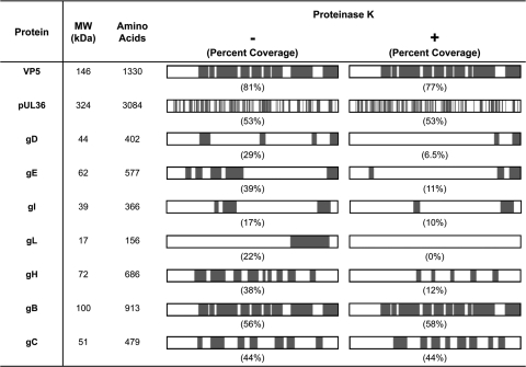
Fig. 6.
Peptide sequence coverage of viral proteins in untreated and proteinase K-treated virions. Schematic representations of each protein are oriented with the N terminus on the left and the C terminus on the right. Regions of the viral proteins that correspond to the peptides we detected by mass spectrometry are indicated in gray, and the percent sequence coverage (by amino acids) is listed in parentheses. The major capsid protein VP5 and the inner-tegument protein pUL36 are contained within the envelope and are therefore predicted to be resistant to proteinase K treatment. The envelope glycoproteins gD, gE, gI, gL, gH, gB, and gC are all predicted to contain domains that are exposed on the virion surface and potentially available for protease cleavage.

