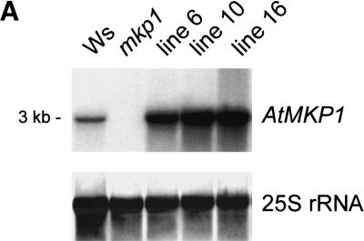
Figure 2.
Expression of the AtMKP1 gene and organization of the mkp1 locus. (A) RNA gel blot analysis of RNA isolated from 3-week-old seedlings. The blot was sequentially probed with AtMKP1 and 25S (Kiss et al. 1989) as a loading control. Ws: Wassilewskija wild type; mkp1 mutant; lines 6, 10, and 16: lines obtained by the transformation of the mkp1 mutant with the genomic fragment containing the AtMKP1 gene. (B) Structure of the AtMKP1 gene and position of the T-DNA insert. Exons are shown as gray boxes. In mkp1, the T-DNA is inserted in the second exon (T-DNA is not drawn to scale). Translational start ATG and the TAA stop codon are indicated. The depicted genomic clone was used for complementation of the mkp1 mutant phenotype in lines 6, 10, and 16.


