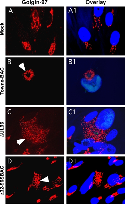
Fig. 4.
Localization patterns of golgin-97 (red) in HF mock transfected (A and A1) or transfected with TowneBAC (B and B1), ΔUL96BAC (C and C1), and Δ32-96SBAC (D and D1) bacmids and fixed at 10 dpt for IFA. Infected cells (arrowheads) were identified by the fluorescence of virally encoded eGFP (not shown). The overlay images (A1, B1, C1, and D1) also include Hoechst 33258 staining, which identifies the nucleus (blue). Original magnification, ×1,000.

