Fig. 5.
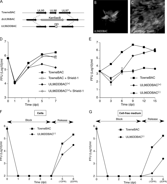
Characterization of a conditional UL96 mutant virus. (A) Schematic of the genome structure of UL96DDBAC (bottom), constructed from ΔUL96BAC (middle), indicating the location of the FKBP-DD relative to the native locus (top). (B and C) eGFP+ plaque phenotype of UL96DDBAC upon transfection into HF at 10 dpt in the absence (B) or presence (C) of 1 μM Shield-1 in the medium. (D) Single-step growth curves of TowneBAC and UL96DDBAC(+) viruses recovered from cells cultured in the presence or absence of 1 μM Shield-1. HF were infected (MOI of 3.0), total viruses (cells plus medium) were harvested at the indicated times postinfection, and titers were determined on HF monolayers without Shield-1. eGFP+ FFU were counted at 10 dpi. The viral titers represent the averages of triplicate experiments, and standard errors are within the symbols. The data point at 0 dpi indicates the input virus dose. (E) Multiple-step growth curves of TowneBAC, UL96DDBAC(+), and UL96DDBAC(−) viruses. HF were infected (MOI of 0.01), total viruses (cells plus medium) were harvested at the indicated times postinfection, and titers were determined and plotted as described above. (F and G) BDCRB block release experiments were performed as described in Materials and Methods. The titers of total cell-associated (F) and cell-free medium-associated (G) viruses were determined and plotted as described above.

