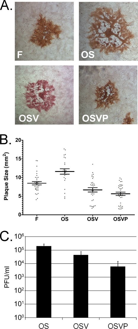
Fig. 2.
Plaque morphology and growth characteristics of OS OSV and OSVP. (A) Nearly confluent Vero cell monolayers were infected with wild-type HSV-1(F), OS, OSV, or OSVP viruses at MOI of 0.001. Individual viral plaques were visualized 48 h postinfection by immunohistochemistry and photographed at the same magnification with a phase-contrast microscope. (B) A minimum of 30 plaques for each virus (prepared as described in the legend to panel A) were measured, and individual plaque sizes are indicated on the x axis, with the horizontal bar indicating the means for each group. Error bars indicate standard error of the means. All pairwise comparisons of the means using two-tailed t tests indicated significance (P < 0.0185) except for the comparison between OSV and OSVP (P = 0.1473). (C) Nearly confluent 4T1 cell monolayers were infected with the above-mentioned viruses at an MOI of 5. Triplicate culture supernatants were collected at 48 hpi, and viral titers were determined (PFU/ml).

