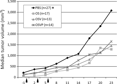
Fig. 4.
Intratumor treatment with OS, OSV, or OSVP viruses. A total of 1 × 10⁵ viable 4T1 cells were implanted subcutaneously in the interscapular area of BALB/c mice. Tumors were measured using a digital caliper at defined time intervals prior to and after treatment (x axis). Median tumor volumes over time are shown on the y axis. Tumors were injected with each virus (1 × 10⁷ PFU/ml in PBS buffer), or PBS alone when tumors reached approximately 80 to 90 mm3 in volume. Tumor volumes were measured prior to (negative values on the x axis) and after the injections. Tumors were treated with each virus at days 1, 3, and 6, as indicated by arrows. The asterisk indicates statistical significance by nonparametric analysis (31). The results shown are from one of three independent experiments that produced similar results.

