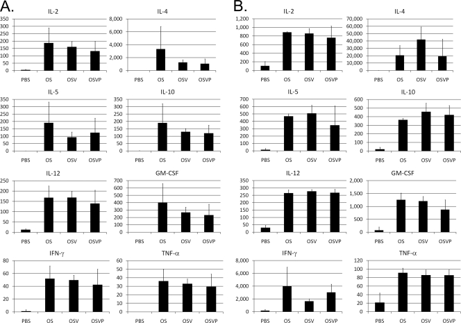
Fig. 7.
Cytokine immunoprofiles of lymphocytes derived from treated mice. Mice with similarly sized 4T1 tumors (200 to 300 mm2) were treated twice with a 3-day interval. Two days following treatments with OS, OSV, OSVP, or PBS (control), lymphocytes from draining lymph nodes were isolated and cultured for 24 h alone (A) or with immobilized anti-CD3 stimulatory antibody (B) at a concentration of 100,000 cells per ml. TNF-α, tumor necrosis factor alpha. Supernatants were assayed for TH1/TH2 cytokine production by Bioplex (Bio-Rad). Shown are the mean quantities of the indicated cytokines in pg/ml with error bars representing 95% CI (n = 3). Shown are results from one of two experiments with similar results.

