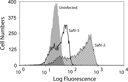
Fig. 6.
FACS analysis of hyperimmune ascitic fluid samples raised in SAFV-2-injected mice and incubated with SAFV-2- or SAFV-3-infected HeLa cells. HeLa cells infected at an MOI of 5 were harvested at 16 h p.i. and stained with a 1:500 dilution of hyperimmune ascitic fluid sample and 1:50 dilution of fluorescein isothiocyanate (FITC)-conjugated goat anti-mouse IgG. The small peaks of SAFV-2 and SAFV-3 overlapping the uninfected cell profile were probably cells that were not infected.

