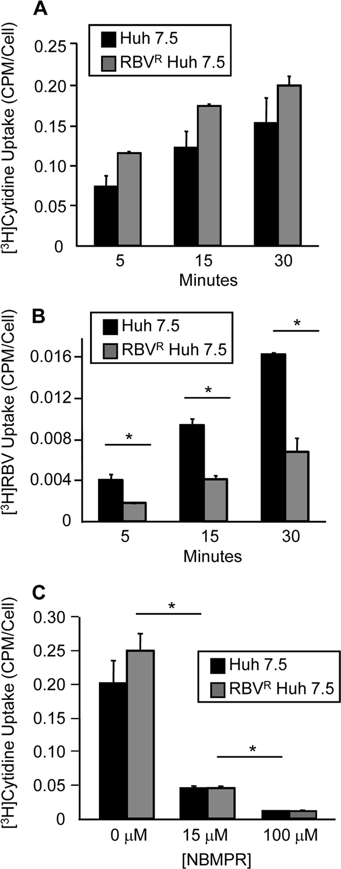
Fig. 3.

Cytidine transport in RBVR and RBVS Huh7.5 cells. (A) Cytidine uptake assay. Cells were incubated with 3H-labeled cytidine, and uptake was quantified at 5, 15, and 30 min postincubation by scintillation counting. The average from two experiments is shown as the number of cpm/cell. (B) RBV uptake was determined as previously described at 5, 15, and 30 min. The average from two experiments is shown. (C) Inhibition of endogenous cytidine uptake. Cells were pretreated with NBMPR, followed by the cytidine uptake assay. Error bars for all panels represent SD, with significance determined using unpaired, two-tailed Student t tests (P < 0.05; Student's t test).
